Vitronectin (VTN or VN) is a glycoprotein of the hemopexin family which is abundantly found in serum, the extracellular matrix and bone.[5] In humans it is encoded by the VTN gene.[6][7]


Vitronectin binds to integrin alpha-V beta-3 and thus promotes cell adhesion and spreading. It also inhibits the membrane-damaging effect of the terminal cytolytic complement pathway and binds to several serpins (serine protease inhibitors). It is a secreted protein and exists in either a single chain form or a clipped, two chain form held together by a disulfide bond.[6] Vitronectin has been speculated to be involved in hemostasis[8] and tumor malignancy.[9][10]
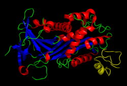
Structure
Vitronectin is a 54 kDa glycoprotein, consisting of 478 amino acid residues. About one-third of the protein's molecular mass is composed of carbohydrates. On occasion, the protein is cleaved after arginine 379, to produce two-chain vitronectin, where the two parts are linked by a disulfide bond. No high-resolution structure has been determined experimentally yet, except for the N-terminal domain.
The protein consists of three domains:
- The N-terminal Somatomedin B domain (1-39)
- A central domains with hemopexin homology (131-342)
- A C-terminal domain (residues 347-459) also with hemopexin homology.
Several structures has been reported for the Somatomedin B domain. The protein was initially crystallized in complex with one of its physiological binding partners: the Plasminogen activator inhibitor-1 (PAI-1) and the structure solved for this complex.[11] Subsequently two groups reported NMR structures of the domain.[12][13]
The somatomedin B domain is a close-knit disulfide knot, with 4 disulfide bonds within 35 residues. Different disulfide configurations had been reported for this domain[14][15][16] but this ambiguity has been resolved by the crystal structure.[16]
Homology models have been built for the central and C-terminal domains.[16]
Function
The somatomedin B domain of vitronectin binds to plasminogen activator inhibitor-1 (PAI-1), and stabilizes it.[11] Thus vitronectin serves to regulate proteolysis initiated by plasminogen activation. In addition, vitronectin is a component of platelets and is, thus, involved in hemostasis. Vitronectin contains an RGD (45-47) sequence, which is a binding site for membrane-bound integrins, e.g., the vitronectin receptor, which serve to anchor cells to the extracellular matrix. The Somatomedin B domain interacts with the urokinase receptor, and this interaction has been implicated in cell migration and signal transduction. High plasma levels of both PAI-1 and the urokinase receptor have been shown to correlate with a negative prognosis for cancer patients. Cell adhesion and migration are directly involved in cancer metastasis, which provides a probable mechanistic explanation for this observation.
| This article uses material from the Wikipedia article Metasyntactic variable, which is released under the Creative Commons Attribution-ShareAlike 3.0 Unported License. |